A dark fleck at the bottom of a vial, or a speck drifting in a freshly drawn syringe, is an unsettling sight during medication preparation. In most cases it is coring: a fragment of the rubber vial stopper sheared off by the needle on the way in. Attention to it is growing. In 2025 the American Society of Retina Specialists (ASRS) published a safety report after members reported cored stoppers in drug vials, and MYCO Medical has fielded more customer questions about stopper fragments in recent months. This guide covers why it happens, how to spot it, and what to change when it does.
What Coring Is and Why It Matters
Coring occurs when a needle passing through an elastomeric vial closure cuts out a small piece of it. The fragment can drop into the vial, lodge inside the needle, or travel into the syringe. Reported incidence ranges from roughly 3 to 40 percent across the studies the ASRS reviewed, because stopper material, needle size and design, and puncture angle all play a part.
Complications reported in the literature include occlusion of venous access devices, pulmonary infarction and granulomas, neurologic sequelae from paradoxical embolism, and anaphylaxis in patients with latex hypersensitivity. Regulators treat any visible particle as grounds to reject a dose: U.S. Food and Drug Administration (FDA) guidance on visible particulates states that injectable units showing visible particles must be rejected, and United States Pharmacopeia (USP) General Chapter <790> lists elastomeric material among the particulates that visual inspection is meant to catch.
The harder problem is that cores are often invisible. In a study of prednisolone acetate vials punctured with an 18G needle, 21 of 200 draws produced a core, and only 11 of those fragments could be seen in the filled syringe. The rest were found only after the syringe was disassembled and rinsed.
Why Coring Happens
No single factor explains coring. The research points to several that stack.
- Needle bore. In a 2022 study of multi-dose propofol vials, 18G needles produced far more rubber particles than 20G or 21G needles regardless of puncture angle. The authors concluded that large bore was the dominant factor.
- Bevel orientation and repeat punctures. A 2023 review in the Indian Journal of Anaesthesia reports more fragmentation when the bevel faces down and the most on a second penetration, whatever the needle size. Multi-dose vials entered repeatedly carry more risk than single-dose vials.
- Stopper thickness and composition. The ASRS report notes that thicker stoppers core more readily than thinner ones and that stopper composition and manufacturing variation also influence coring. This matters when a drug maker changes vial suppliers or stopper formulations. As packaging manufacturer West explains in its discussion of USP <382> fragmentation testing, a closure is qualified for a specific needle and puncture procedure, so a different closure can behave differently under the same technique. A routine that has never cored can begin producing fragments with no change in the needle at all.
- Needle design. Design matters, and not always in the expected direction. A 2014 study found coring in 40.8 percent of vials entered with a blunt plastic cannula versus 4.2 percent with an 18G sharp steel needle. Steel blunt fill needles are a different device, but a 2022 case report documents a core drawn through an 18G steel blunt fill needle too, so no drawing-up needle is immune. Entry angle also matters, although the findings are mixed: a 2015 study of propofol vials found that angled puncture reduced coring with a blunt needle compared with perpendicular puncture, while the propofol study above found the opposite with an 18G sharp needle, which suggests the sliding, bevel-up motion matters more than the angle alone. The RELI® Blunt Fill Needle is built around that principle, with an angled bevel designed to reduce coring of the vial stopper and a 1.5-inch length that aids penetration of different septum barriers.
How to Identify Coring
Inspection is the first line of defense, at three points.
- Inspect the vial after needle entry. Look for a dark fleck at the base of the vial or a divot in the stopper. In the ASRS cases, clinicians noticed the particle in the vial rather than the syringe.
- Inspect the drawn syringe and the needle. Hold the syringe against black and white backgrounds under good light and rotate it slowly, the method USP <790> describes. A fragment can also sit inside the needle or hub, so check the drawing needle after removal.
- Assume you can miss it. Because roughly half of the cores in the prednisolone study were not visible in the syringe, inspection alone cannot clear a dose. Filtration closes that gap.
How to Prevent Coring
Technique comes first because it costs nothing to change.
- Enter at an angle, bevel up. The method described in Anesthesia & Analgesia is to orient the bevel up, start at roughly 45 to 60 degrees, apply slight lateral pressure so the bevel slides through the elastomer rather than punching it, and bring the needle to vertical only as the bevel clears the stopper.
- Use the smallest bore that will do the job. An 18G needle remains the standard for viscous medications and large volumes, but a 20G needle draws most aqueous solutions comfortably with less rubber at risk.
- Puncture once. Avoid re-entering the same stopper, and prefer single-dose vials where practical.
Device choice is the second layer.
- Use a filter needle when particulate is unacceptable. In one experimental study, aspirating through an 18G needle fitted with a 5-micron filter eliminated rubber particles entirely, while unfiltered 18G and 23G draws produced particles in 10 and 33 percent of samples. The ASRS report likewise recommends steel filter needles for drawing up single-dose vials. The RELI® Blunt Fill Needle with Filter, Sterile, 18G x 1.5 inch (SKU F-BFN18G151) combines the angled blunt bevel with an integrated filter for this purpose.
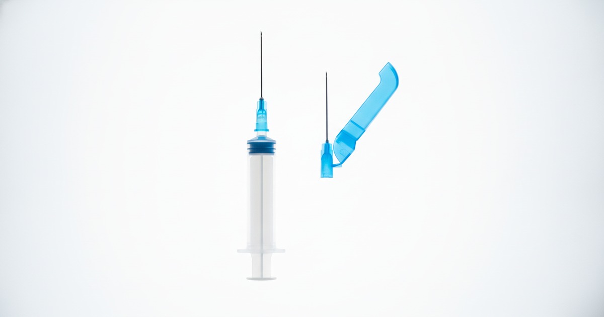
- Reduce handling steps. The RELI® Blunt Fill Needle with Syringe pairs an 18G x 1.5-inch blunt needle with a pre-attached 3, 5, or 10 mL syringe, removing an assembly step.
- Swap the drawing needle before administration. A fresh injection needle keeps any fragment trapped in the drawing needle or filter from reaching the patient. Removing the sharp from the drawing-up step has its own safety case, covered in our guide to reducing sharps injuries in hospitals.
Troubleshooting When Coring Occurs
- Stop and discard. Do not administer the dose or try to fish the fragment out. The ASRS report advises discarding the medication in a cored vial. Prepare a fresh dose from a new vial with a new needle and syringe.
- Record the details. Note the drug, lot number, vial size, needle gauge, and type, and how the stopper was entered. Retain the components if policy allows.
- Look for the pattern before blaming the device. If fragments follow one drug or vial lot across different needles, the closure is the likely variable, and it is worth asking whether the vial supplier recently changed. If they follow one needle type across many vials, the device or technique is.
- Report it. FDA guidance calls for investigation and corrective action when visible particulates appear. Report suspected defects to the manufacturer and through MedWatch, and log the event internally.
- Then adjust. That may mean a smaller gauge, retraining on angled entry, single-dose vials, or adding a filter needle to the preparation kit.
Explore RELI® Medication Preparation Solutions
MYCO Medical supplies blunt fill, blunt filter, and pre-attached syringe configurations as part of its needles and syringes line. If your teams are seeing stopper fragments, contact our sales team to request evaluation samples of the filter and standard blunt fill configurations to test against your current vials.
Frequently Asked Questions
What is vial coring?
Coring is the shearing of a small piece of a vial rubber stopper by the needle during puncture. It can end up in the vial, the needle, or the syringe, and is often too small to see.
Do blunt fill needles cause coring?
Any needle that passes through a rubber stopper can core it. Blunt plastic cannulas cored far more often than sharp steel needles in one study, while steel blunt fill needles with an angled bevel are designed to reduce coring. Gauge, technique, and the stopper itself matter as much as the device.
Why has coring started with vials we have used for years?
Stopper thickness, composition, and manufacturing variation all influence coring, and drug makers change vial suppliers and stopper formulations. If fragments track a particular drug or lot rather than a needle, the closure is the most likely explanation.
Does entering at a 45-degree angle prevent coring?
It helps as part of a bevel-up, sliding entry that finishes vertically. Angle alone is not enough: one study found an 18G needle at 45 degrees cored more than at 90 degrees, so gauge, single puncture, and filtration matter as well.
Can a cored dose be used if it is filtered?
No. Discard the dose and prepare a fresh one from a new vial. Filter needles are a preventive tool used during the draw, not a rescue for a contaminated syringe.
Should coring be reported?
Yes. Log it internally with the drug, lot, and device details, notify the drug manufacturer, and report suspected defects through FDA MedWatch. Patterns across lots reveal the root cause.


.jpg)

%20(1).jpg)
%20(2).jpg)